Electron Microscope: Scanning Electron Microscope, Hitachi
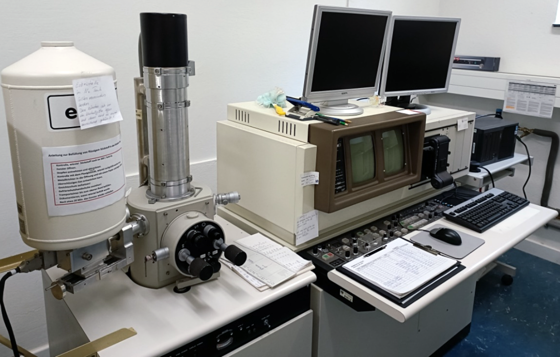
The electron microscope uses electron radiation to produce a magnified image of objects for the determination of surfaces and/or the element composition. The images of the object surfaces have a high depth of field.
- General description: scanning electron microscope with elemental Analysis (EDX) (see electron microprobe analysis (EMPA))
- Application aim: examination of very small objects in different magnifications and simultaneous analysis of the elements present and their quantity
- Mobility: not mobile
- Equipment specifics:
- Magnification up to 4000x
- Tungsten wire serves as the cathode
- 3D view of objects
- EDX for elemental analysis
- Application requirements: none
- Sample required: yes, only small samples
- Contact required: no
- Interaction spot dimensions: different, depending on the magnification
- Limitations: frequent sample change, since only one sample can be examined at a time; damage to the objects by the electron beam; the objects to be examined must be dry; out of focus at higher resolutions
- Set up time: approx. 30 minutes
- Average time for measuring: image capture immediately; for EDX measurements 5-10 minutes
- Average time for processing: 1 minute
- Software package: WINEDS V4.0 and DIPS (Digital Image Processing System 2.6)
- Output: .tif for images; EDX-results as .csv and .bmp files
- Contact: Jörg Fromm, joerg.fromm"AT"uni-hamburg.de
- Location of the equipment: Institute of Wood Science (Leuschnerstr. 91 d, Hamburg)