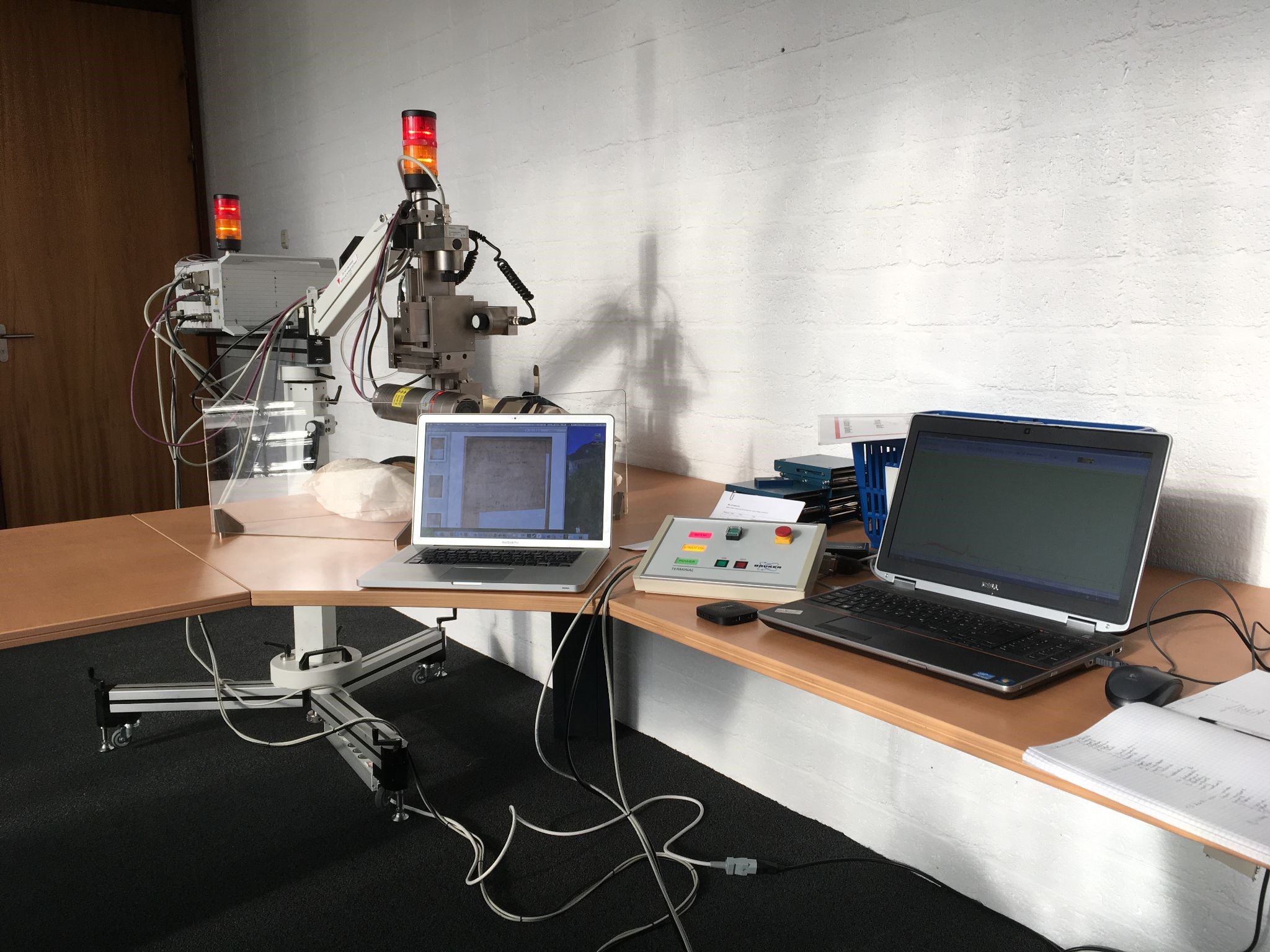
XRF-Spectrometer; Artax, Bruker Nano GmbH

CSMC
This XRF-spectrometer is well known in the field of cultural heritage and is standard equipment in the majority of large museums. It has a measuring spot size of 70 µm diameter, a CCD camera for sample positioning, and an electrothermally cooled Xflash detector (SDD, area: 30 mm2) with an energy resolution of <150 eV at 10 kcps. The movable probe is operated by XYZ motors that allow for spot measurements as well as line and small area scans. Open helium purging in the excitation and detection paths allows for detection of light elements (Z = 11). Measurements are made using a 30 W low-power Mo tube, operating at 50 kV and 600 µA, with an acquisition time of 10-100 s (live time). The mobile XRF probe moves over the object at a distance of 5 mm and stops for the duration of a single measurement. The areas of investigation are usually determined beforehand. This instrument is used for a quantitative analysis of iron gall inks, pigments, etc.
- General description: XRF-spectrometer; no preparation of the sample required, non-destructive
- Application aim: elemental analysis
- Mobility: transportable device; can be packed in 4 boxes for transport (about 150 kg total when in boxes)
- Equipment specifics:
- X-ray source: air-cooled Mo X-ray tube
- 10-50 kV, 5-600 μA, 30 W
- Laser assisted focus, with built-in camera
- electro-thermally cooled X-flash detector (SDD, area: 10 mm2) detector
- Application requirements: controlled temperature, 18-25 oC and relative humidity ≤ 80%, safety requirements (X-rays)
- Sample required: no, surface to analyse needs to be somewhat flat
- Contact required: no
- Interaction spot dimensions: ~ 100 µm
- Limitations: limited equipment mobility, no mapping capacity
- Set up time: ca. 30 min for warming up the tube and calibration against a standard metallic alloy plate made of copper and zinc
- Average time for measuring: 5-10 min per linescan + set up time
- Average time for processing: about 5-10 min per spectrum
- Software package: Windows 7, Bruker Spectra
- Output: .rtx (for linescans) or .spx (for individual spectra ~15KB)
- Contact: Sebastian Bosch, sebastian.bosch"AT"uni-hamburg.de
- Location of the equipment: CSMC Lab (Warburgstr. 28, Hamburg)
