Raman-Spectrometer; Renishaw inVia Raman Spectrometer
CSMC
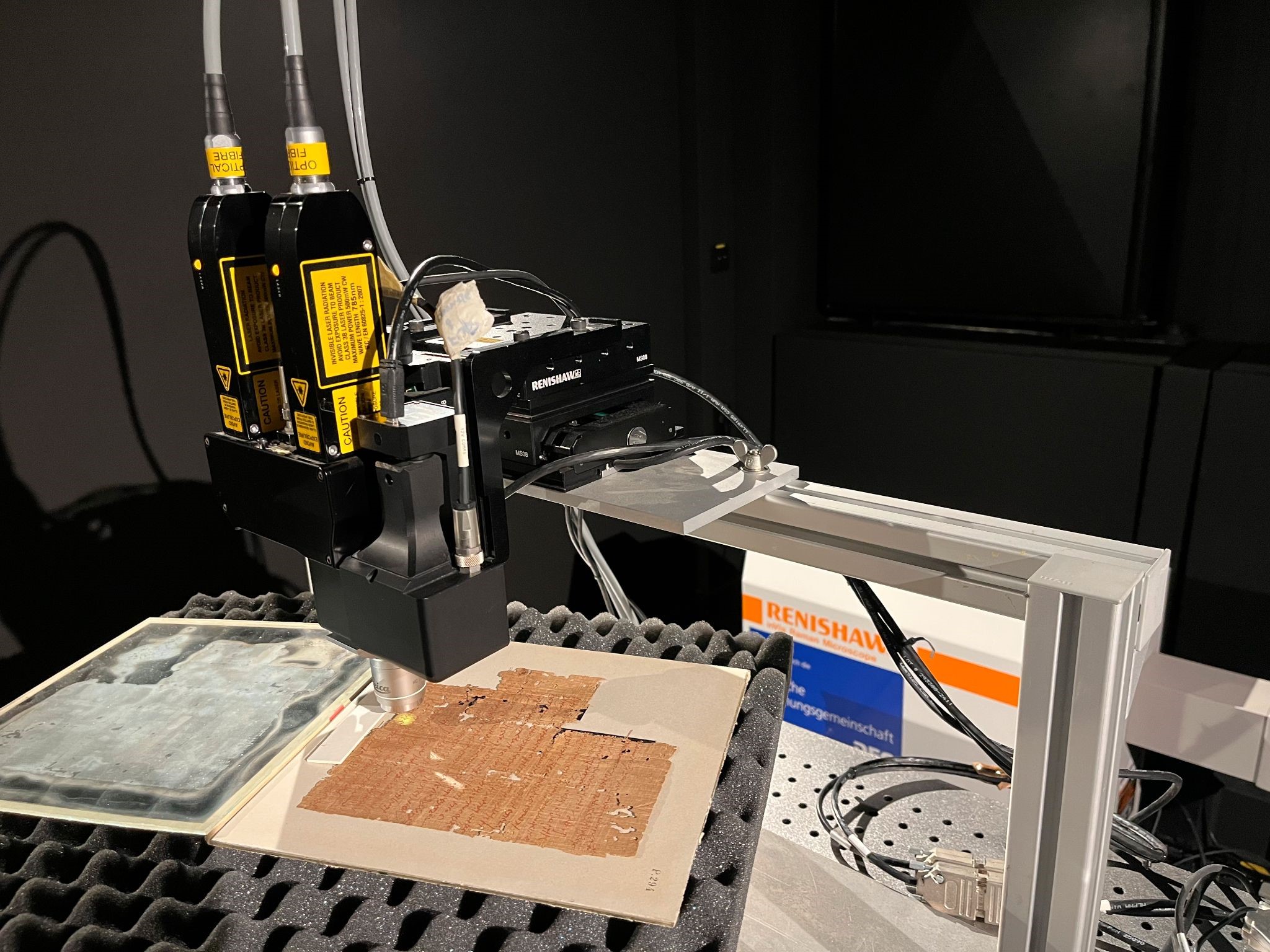
The In Via Raman spectrometer has been specially adapted for the study of objects in the Cultural Heritage field. It is equipped with two fiber optics probes connected to lasers operating at 532 and 785 nm, respectively. The probes are connected to a camera to position the object and a CCD camera for signal registration. Measurements can be carried out with a lens of x20, x50 and x100 magnification and an output power of 5-10 mW in the spectral range 100-3600 cm-1 with a spectral resolution of 4 cm-1.
- General description: Renishaw inVia Raman spectrometer (specially adapted for the study of objects in the Cultural Heritage field).
- Application aim: Determination of the vibrational modes of crystalline or amorphous phases, the resulting spectrum is a structural fingerprint by which molecules can be identified. This spectrometer is used mostly to identify pigments and inks.
- Mobility: stationary device; ca. 250 kg in total (including the spectrometer, lasers, fibre probe, XYZ stage, and optical table)
- Equipment specifics:
- Peltier-cooled charge-coupled detector (CCD array, 1024 x 248 pixel): spectral range: 100 - 3600 cm-1, spectral resolution: 4 cm-1, instrumental peak position accuracy: 0.35 cm-1
- Two different lasers for excitation: 785 nm diode laser: 300 mW, grating with 1200 lines/mm, 532 nm Nd:YAG laser: 100 mW, grating with 1800 lines/mm
- Measuring head (dual fibre optical probe) with x20, x50 and x100 Long-Working Distance (LWD) objectives
- XYZ stage (25 mm travel distance)
- Application requirements: dark room, vibration-free environment, controlled temperature, safety regulations (laser class 3B), 8-25 °C and relative humidity ≤ 80%
- Sample required: no, surface to analyse needs to be flat, no preparation of the sample required, non-destructive, analysis of small details possible (thanks to the microscope objectives)
- Contact required: no
- Interaction spot dimensions: ~10 μm (for x100 objective), 20 μm (for x50 objective),
50 μm (for x20 objective) - Limitations: competition with fluorescence on some samples
- Set up time: ca. 30-40 min for warming up the laser and calibration against a silicon standard wafer (peak at 520 cm-1)
- Average time for measuring: from a few seconds up to 5 minutes per spot, + set up time
- Average time for processing: from a couple of min/spectrum for quick identification to 15-30 min (depending on the complexity of operations required like baseline subtraction and peak fitting)
- Operating system: Windows 7
- Software package: WIRE
- Output: .wxd (proprietary format of Renishaw, ~150 KB) or .txt (~10 KB) or .spc (~10 KB)
- Evaluation program: OriginPro 2019b, OMNIC, OPUS
- Contact: Sebastian Bosch, sebastian.bosch"AT"uni-hamburg.de
- Location of the equipment: CSMC Lab (Warburgstr. 28, Hamburg)